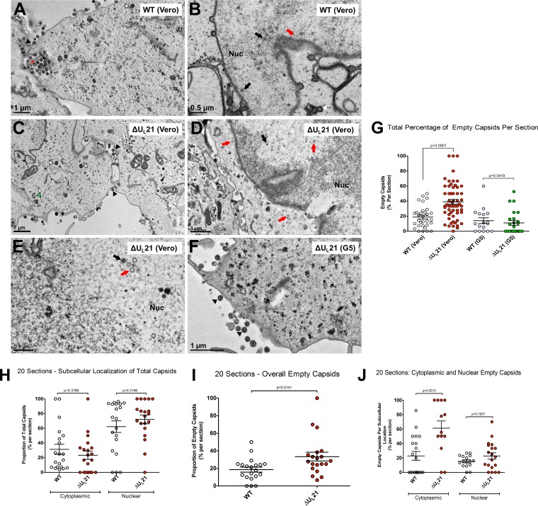
FIG 3.
Morphology of capsids produced by the UL21 deletion virus. (A to F) Vero or G5 cells were infected at an MOI of 10 with either wild-type or ΔUL21 virus. The samples were fixed and processed for electron microscopy at 24 h postinfection, and representative images are shown. The black arrowheads (A, C, and F) mark mature extracellular particles. The red asterisk (A) marks a rare enveloped extracellular empty capsid. The black arrows (B, D, and E) indicate complete, DNA-filled capsids, whereas the red arrows (B, D, and E) denote empty capsids. The green arrowhead (C) marks a partially wrapped capsid in the cytoplasm, and the white arrowheads (A and C) show wrapped capsids in the cytoplasm. Where applicable, the nucleus (Nuc) is indicated. (G) The percentage of empty capsids within each total capsid population, regardless of subcellular position, was plotted for the wild-type and deletion viruses on Vero or G5 cells, and the error bars indicate means and SEM. One-way ANOVA revealed significant differences in the percentages of empty capsids among the groups, and the relevant P values (from unpaired t tests) are shown. (H to J) For analysis of the subcellular locations of empty capsids produced by the wild-type and mutant viruses, a subset of 20 independent sections, all having the nuclei and cytoplasm in view, were assessed as described for panel G and are explained in the text.

