FIG 1.
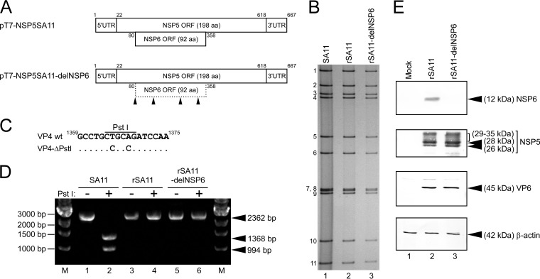
Generation of the rSA11-delNSP6 virus incapable of expressing the NSP6 protein. (A) Schematic presentation of the plasmids encoding segment 11 (NSP5/6 genes), used for rescue of the wild-type (wt) rSA11 and mutant rSA11-delNSP6 viruses (pT7-NSP5SA11 and pT7-NSP5SA11-delNSP6, respectively). To generate plasmid pT7-NSP5SA11-delNSP6, the start codon and three AUG codons in the NSP6 ORF of SA11 were disrupted by replacing them with ACG codons. The arrowheads indicate the ACG mutation sites. UTR, untranslated region; aa, amino acid. (B) PAGE of viral genomic dsRNAs extracted from the native SA11, rSA11, and rSA11-delNSP6 viruses. Lane 1, dsRNAs from the native SA11; lanes 2 and 3, dsRNAs from the rescued rSA11 (lane 2) and rSA11-delNSP6 (lane 3) viruses. The numbers on the left indicate the order of the genomic dsRNA segments of the native SA11. (C) Rescued rSA11 and rSA11-delNSP6 contain a signature mutation in their VP4 genes as a gene marker. Nucleotide substitutions (T to C and A to C at nucleotide positions 1365 and 1368, respectively) were introduced to eliminate a unique PstI site in the VP4 gene (15). (D) The VP4 gene RT-PCR products were digested with PstI, followed by separation in a 1.2% agarose gel. Native SA11 (lanes 1 and 2), rSA11 (lanes 3 and 4), and rSA11-delNSP6 (lanes 5 and 6) are shown. The 2,362-bp fragments (lanes 1, 3, and 5) were digested with PstI (lanes 2, 4, and 6). M, 1-kb ladder markers. (E) Expression of the NSP6 protein and other RVA proteins in MA104 cells infected with rSA11 or rSA11-delNSP6. Whole-cell lysates of infected cells were analyzed by immunoblotting using anti-NSP6 antiserum, anti-NSP5 antiserum, anti-VP6 antiserum, or anti-β-actin monoclonal antibody. The anti-β-actin antibody was used as a loading control. Mock (lane 1), rSA11 (lane 2), and rSA11-delNSP6 (lane 3) virus infection is shown.

