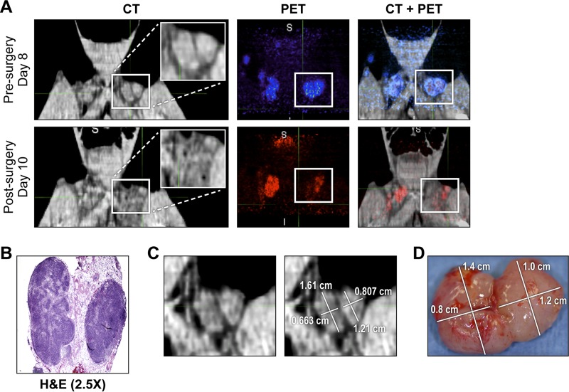
FIG 4.
Tracking removal of inguinal LNs by PET/CT imaging. (A) CT, PET, and fused PET/CT images of the inguinal LN area in the untreated survivor (UNTR-2) acquired on days 8 and 10 p.i., before and after LN biopsy (day 9 p.i.), respectively. On the CT image, a white box marks the left-side inguinal LNs with an inset image of the magnified LN area. On day 8 or 10 p.i., the PET signal within the inguinal LN area was color-coded blue or red, respectively. (B) H&E staining of day-9-p.i.-biopsied LNs. (C) CT image of the two left-side LNs acquired on day 8 p.i. The sizes of the two LNs were determined using MIM5 software. (D) The actual sizes of the LNs were determined after removal on day 9 p.i. Abbreviations: H&E, hematoxylin & eosin; LN, lymph node; p.i., postinfection; UNTR, untreated.

