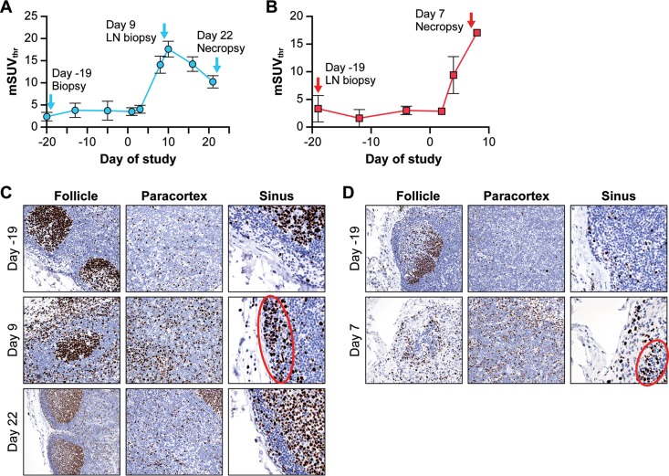
FIG 5.
LN tissue characterization by immunohistochemistry and PET/CT imaging. (A) Changes in [18F]-FDG uptake as measured by mSUVthr in axillary LNs of surviving NHPs. (B) Changes in [18F]-FDG uptake measured via mSUVthr in axillary LNs of moribund NHPs. (C) Ki-67 staining of inguinal LN tissues from a surviving NHP (CDV-1) collected on day −19 preexposure and days 9 and 22 p.i. Areas of follicle, paracortex, and subcapsular sinus were scored for Ki-67-positive cells. The red oval shows Ki-67-positive proliferating B cells in the sinus. (D) Ki-67 staining of inguinal LN tissues from a moribund NHP (UNTR-1) collected on day −19 preexposure and day 7 p.i.at necropsy. Areas of follicle, paracortex, and subcapsular sinus were scored for Ki-67 and CD4, CD8, or CD20 (data not shown). The red oval shows Ki-67-positive proliferating B cells in the sinus. Abbreviations: CDV, cidofovir; [18F]-FDG, [18F]-fluorodeoxyglucose; LN, lymph node; mSUVthr, modified SUVthreshold; NHP, nonhuman primate; p.i., postinfection; UNTR, untreated.

