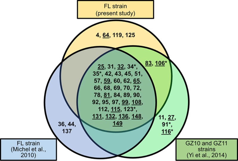
FIG 1.

CyHV-3 virion proteome. Schematic representation of CyHV-3 virion-associated proteins identified in independent studies: upper circle, analyses of the European FL strain performed in the present study; lower left circle, analyses of the FL strain performed in a former study (9); and lower right circle, analyses of two Chinese strains (GZ10 and GZ11) (10). Numbers represent CyHV-3 ORFs. Asterisks indicate viral proteins that were detected in only one of the two Chinese isolates. Predicted transmembrane proteins are underlined.
