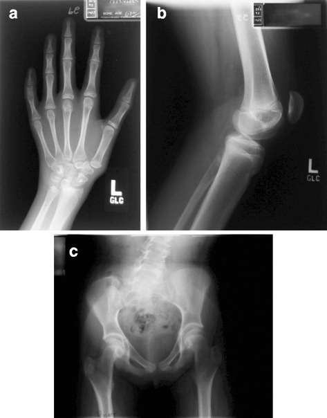
Fig. 2.

a Radiograph of the left hand for bone age: The distal phalanx of each digit is small and narrow. Shortening of the middle phalanx of digits 2 and 5. Slender metacarpals 4 and 5 and short 5th metacarpal. The margins of the carpal bones are more angular than smooth. b Lateral radiograph of the Left Knee: The patellar height ratio, measured length of the patellar tendon divided by length of the patella is 1.7, normal is 1.5. Typically increased PHR is associated with increased risk of patella dislocation. c AP standing pelvis radiograph: Pelvic tilt demonstrating a leg length discrepancy. The iliac bones are narrow. Bilateral Coxa Valga is present. Acetabular coverage is good. Dysplasia of the femoral epiphysis (flat and small)
