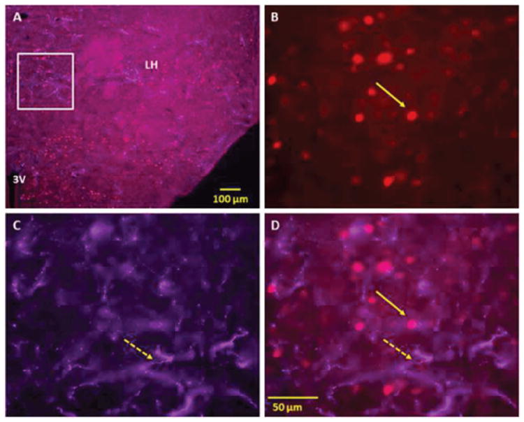
Figure 2.

Double immunofluorescence staining demonstrating the location of orexin (Orex) immunoreactive (IR) nerve fibers with respect to nicotine (NIC) induced c-Fos IR cells of lateral hypothalamus (LH). Panel A: Low power fluorescent image showing NIC-induced c-Fos IR and Orex IR nerve fibers in LH Panels B-D: High power images showing NIC-induced c-Fos IR cells (B), Orex IR nerve fibers (C) and merged images of c-Fos with Orex IR fibers (D). Orex IR nerve fibers are seen in areas overlapping NIC induced c-Fos activated cells of LH and several other hypothalamic regions. Solid arrows point to representative c-Fos IR and broken arrows to orexin IR nerve fibers. Square indicates magnified area (3V 3rd Ventricle).
