
Figure 2. Detection of ROS producing cells in the stromal compartment of Muc2+/+ and Muc2−/− mice.
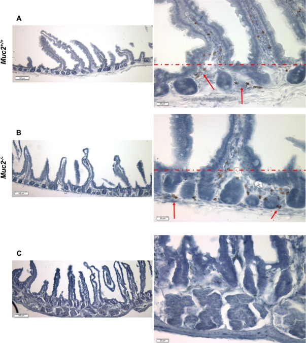
(A–C) show representative micrographs of frozen sections of flat mucosa of small intestine from 3 month old Muc2+/+(A) and Muc2−/− (B) mice, respectively, incubated in vitro with DAB, as described in Material and Methods. Red arrows indicate cell positivity due to DAB polymerization in the presence of cell-generated H2O2. (C) shows loss of signal, and thus specificity of the reaction, upon addition of catalase to the reaction buffer. The red dotted line separates the intravillus area from the pericrypt area. Magnification bars are shown.