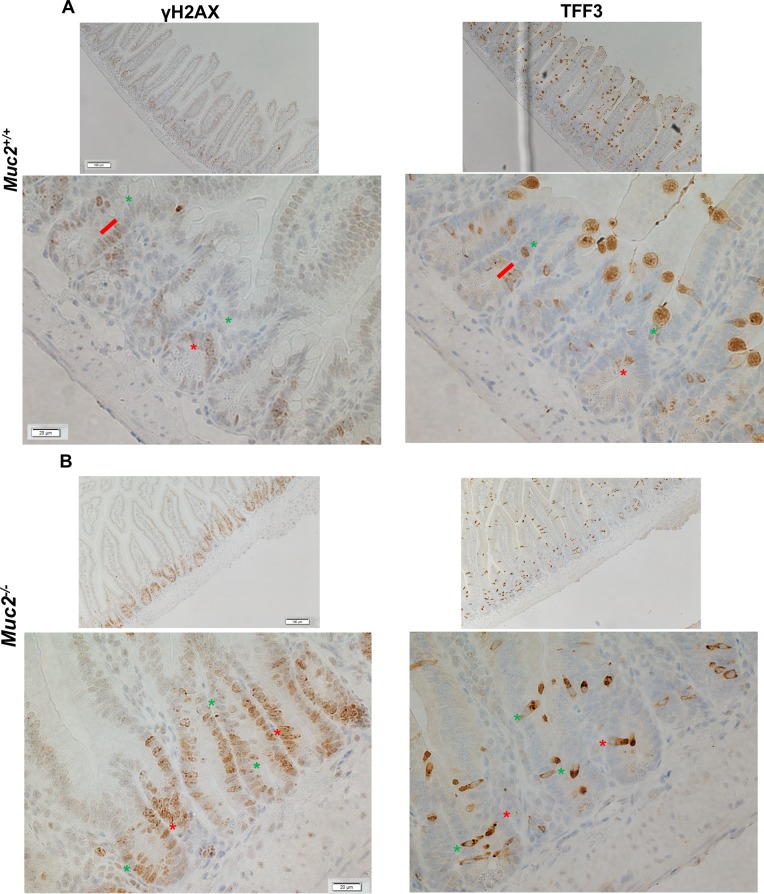
Figure 4. Increased number of γH2AX positive cells in Muc2−/− crypts.
Immunohistochemical detection of γH2AX (left column), a marker for DNA damage, and TFF3 (right column), a marker of goblet cells in serial sections of SI from 3 month old mice of the indicated phenotypes, (A) and (B) for Muc2+/+ and Muc2−/−, respectively. In A and B red lines and asterisks identify γH2AX-positive goblet cells, expressing TFF3. Green asterisks pinpoint at γH2AX-positive cells that do not express TFF3.

