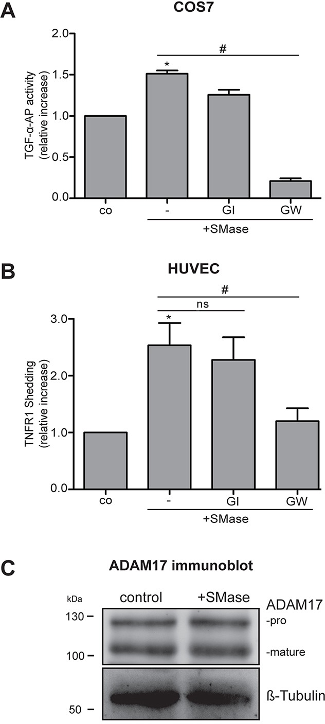
Figure 1. Extracellular SMase induces ADAM17-mediated shedding of TGF-α-AP in COS7 cells and TNFR1 release in HUVECs.

(A) Cells were stimulated with 0.1 U/ml SMase in the presence and absence of the ADAM10 inhibitor GI254023X (GI, 3 μM) or the ADAM17 and −10 inhibitor GW280264 (GW, 3 μM). The release of the transfected AP-tagged TGF-α was determined in COS7 cells upon 3 h of SMase treatment. (B) The release of the endogenous ADAM17 substrate TNFR1 in the supernatant was measured by ELISA in HUVECs after 2 h of SMase stimulation. Values are plotted as relative increase compared to control. * indicates a significant increase compared to control, # indicates a significant reduction in comparison to the stimulated sample, ns: no significant differences (*#: p<0.05; n=5; ± SEM). (C) HUVECs were stimulated with SMase (0.1 U/ml) for 2 h and analyzed for ADAM17 expression by immunoblot analysis. No changes in the amount of mature or pro-ADAM17 were observed. β-Tubulin was used as loading control.
