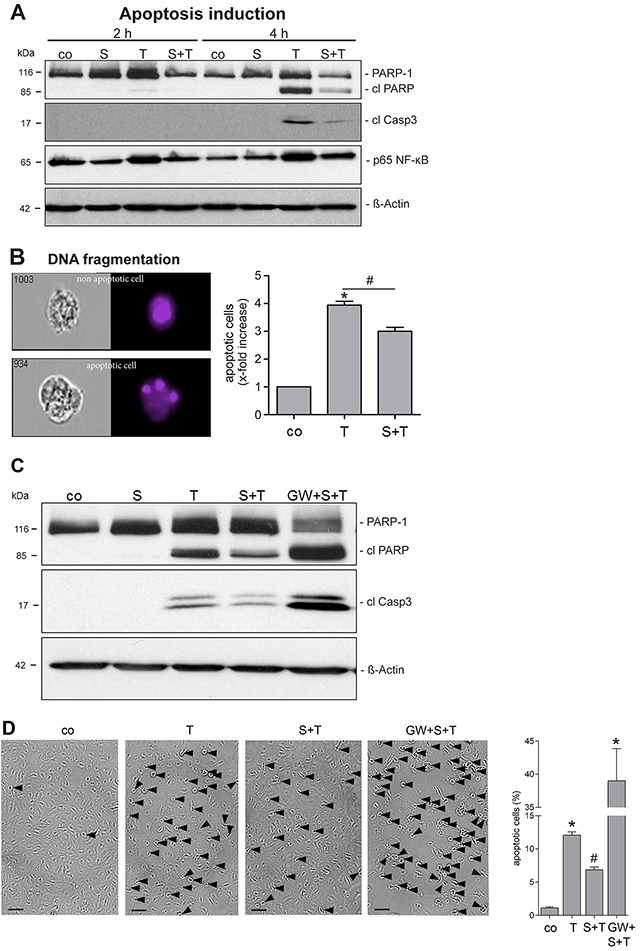
Figure 4. SMase treatment reduces the TNF-α sensitivity of endothelial cells.

(A) HUVECs were pretreated with SMase (S, 0.1 U/ml, 2 h) and stimulated with TNF-α (T, 40 ng/ml + CHX (5 μg/ml) for 2 or 4 h, followed by lysis and Western blot analyses. Shown are representative Western blots for total PARP-1, cleaved PARP (cl PARP), cleaved caspase-3 (cl Casp3), and phosphorylated p65 NF-κB. β-Actin served as a loading control. (B) Apoptosis was additionally measured by quantifying nuclear fragmentation by ImageStream analysis. HUVECs were analyzed after overnight incubation with TNF-α/CHX with or without SMase pretreatment as described. The means of three different independent experiments are shown. * indicates a significant increase compared to control, # indicates a significant reduction in comparison to the stimulated sample (*#: p<0.05). Data are represented as mean ± SEM. (C, D) TNF-α-induced cell death is reduced upon SMase pre-treatment and reinforced upon ADAM17 inhibition. HUVECs were pretreated with SMase (SM, 0.1 U/ml, 2 h) in the presence or absence of the inhibitor GW (3 μM) and stimulated with TNF-α (T, 40 ng/ml) + CHX (5 μg/ml) for 4h. (C) Cells were analyzed by Western blotting. Shown is a representative blot (n=4) for cleaved PARP-1 (cl PARP), and cleaved caspase-3 (cl Casp3), β-Actin served as a loading control. (D) The number of HUVECs showing TNF-α-induced cell membrane blebbing is reduced upon SMase pre-treatment and reinforced upon ADAM17 inhibition. Cells were stimulated as described above and photographs were taken. Scale bars, 50 μm. The percentage of cells was determined by counting blebbing cells compared to the number of total cells on 3 randomly selected fields. * indicates a significant increase compared to control, # indicates a significant reduction in comparison to the TNF-α-stimulated sample (*#: p<0.05; n=6; ± SEM; one-way ANOVA with post-hoc Tukey test).
