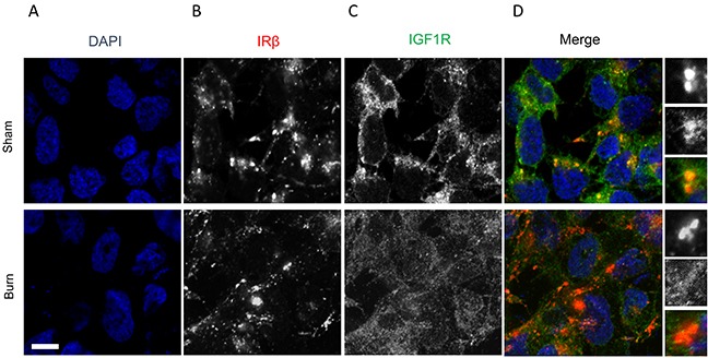
Figure 2. Immunofluorescence assay to detect relative expression of IRβ and IGF1R protein in anterior tibial muscle obtained from burn and sham rats at day 7.

(A) DAPI staining, (B) IRβ, (C) IGF1R, and (D) merge images. Scale bar: 100 μm. Images are representative of at least 10 different images obtained within the experimental set.
