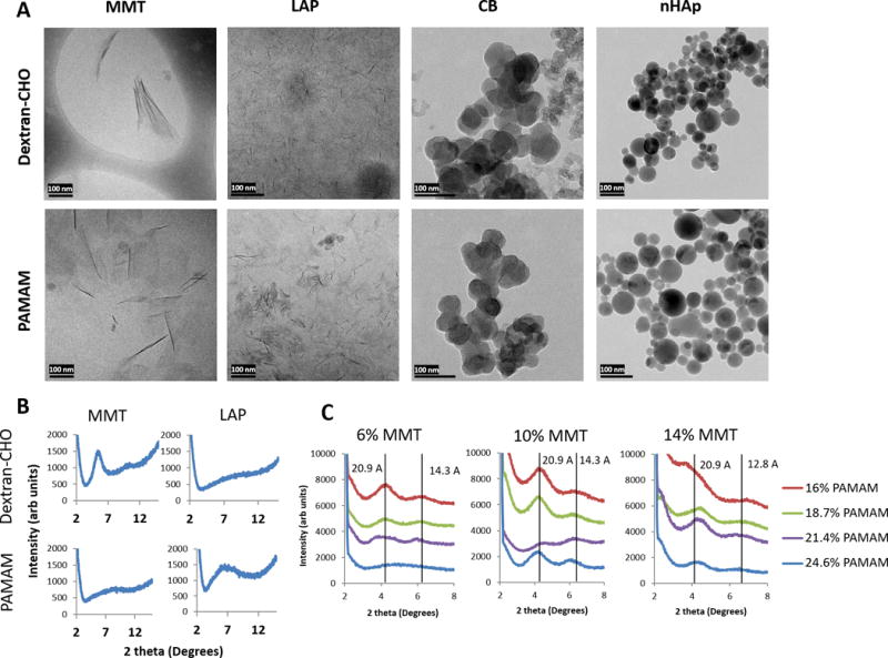
Figure 2.

Morphology and dispersion of nanofillers. (A) Cryo-TEM of dispersions of each of four nanofillers in either 20% dextran-aldehyde or 24.6% PAMAM dendrimer macromer solutions. MMT = montmorillonite, 6%, LAP = Laponite, 6%, CB = carbon black, 5%, nHAp = nanoscale hydroxyapatite, 5%. Every dispersion was diluted 1:100 immediately prior to freezing to reduce background. Scale bars = 100 nm. (B) Representative X-ray diffraction spectra of MMT and LAP nanoplatelets in 20% dextran-aldehyde or 24.6% dendrimer; the presence of a peak indicates nanoplatelet stacking and a lack of complete exfoliation. (C) Varying polymer-nanoplatelet ratios results in varying exfoliation; at high PAMAM concentration and low MMT concentration (6% MMT, 24.6% PAMAM) exfoliation appears complete, but as polymer concentration decreases or MMT concentration increases, increasing XRD signal is indicative of intercalated and tactoid structures. Vertical lines at each peak are labeled with the corresponding d-spacing, giving approximate inter-platelet distances in Angstrom.
