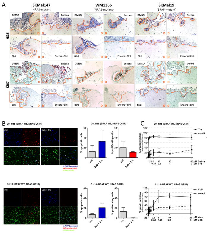
Figure 2. MEKi combined with BRAFi inhibit invasive tumor growth in organotypic skin cultures and lead to apoptosis induction and reduction in proliferation in patient-derived tumor slice cultures.
(A) Melanoma cells were seeded onto organotypic skin reconstructs, incubated for 4 days and then treated for 10 days with DMSO (control) or 0.1 (BRAF-mutant cells) and 1 μM (NRAS-mutant cells) encorafenib or/and binimetinib. Fixed tissue sections were stained with H&E or Ki67. An orange line marks the tumor front. T = tumor cells, D = dermis, Scale bar = 10 μm. (B) Tumor tissue from patients with NRAS-mutant melanoma was expanded in a PDX model, sliced and then treated with the indicated inhibitor combinations. After 4 days the slices were stained with YO-PRO-1 (nuclei marker), Ki67 (proliferation marker) or cleaved PARP (apoptosis marker) and analyzed by confocal microscopy. Images of one representative per treatment group are shown. The percentage of apoptotic and proliferating cells was quantified and displayed in a bar graph (error bars represent SD of 4 random sites in each tumor per treatment group). (C) NRAS-mutant patient-derived melanoma cells were treated with the indicated inhibitors for 72 h. The percentage of growth inhibition was calculated, normalized to the DMSO-treated control (mean ± SD of quadruplicates).

