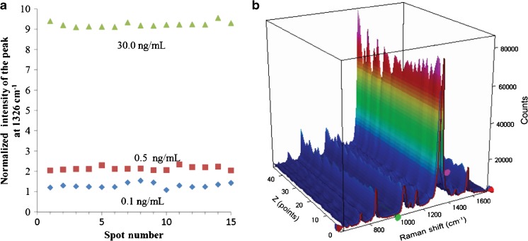
Fig. 8.
a Reproducibility of three separately prepared SERS immunoassays exposed to different concentrations of IL-8 in blood plasma (0.1, 0.5, and 30.0 ng mL−1). SERS spectra were recorded at 15 randomly selected spots on the substrate in each SERS assay. b Representative two-dimensional SERS spectra recorded in SERS assays of 30.0 ng mL−1 IL-8 performed at 40 different spots on the SERS surface. The spectra were collected over a distance of 1 mm in 10 μm steps (40 spectra are shown). Each point on the map was recorded using 5 mW of excitation at 632 nm and an integration time of 10 s

