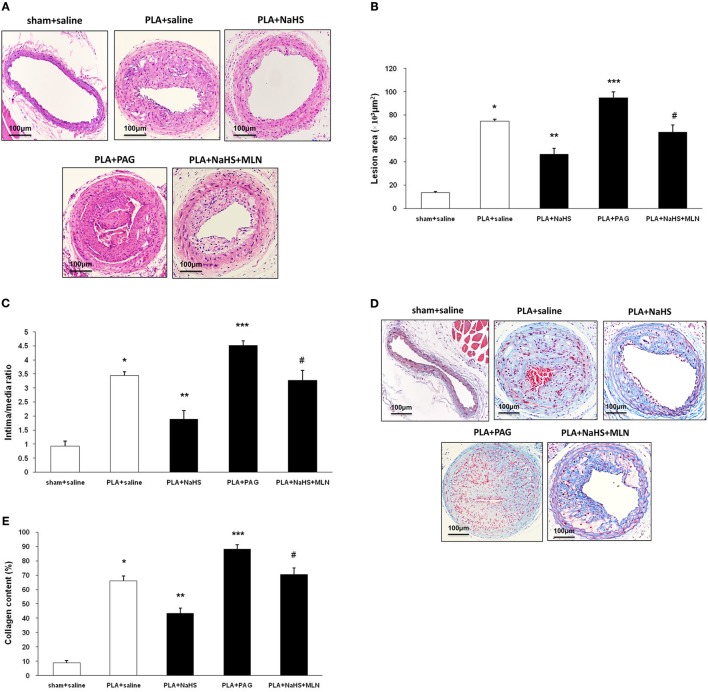
Figure 3.
Effect of NaHS or PAG on PLA-induced atherosclerosis in LCA. Representative light microscopy images of hematoxylin and eosin-stained (A) or Masson trichrome-stained (D) LCA cross sections were taken from sham-operated mice with saline, PLA mice with NaHS, PLA mice with PAG, or PLA mice with NaHS and MLN (intervention with MLN-4760 for 14 days). Scale bar for histological images = 100 μm. Lesion area of intima (B), intima/media ratio (C) and collagen deposits (E) in the neointimal hyperplasia in LCA were measured as described in Materials and Methods. Results shown are the mean ± SEM (n = 6 animals in each group). *P < 0.05 for the comparison between sham+saline and PLA+saline. **P < 0.05 for the comparison between PLA+saline and PLA+NaHS. ***P < 0.05 for the comparison between PLA+saline and PLA+PAG. #P < 0.05 for the comparison between PLA+NaHS and PLA+NaHS+MLN.

