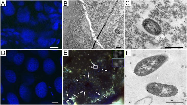
FIGURE 3.

Mollicutes bacteria in A. cephalotes across developmental stages. (A,D) Absence of Mollicutes in larval (A) and pupal (D) tissues of A. cephalotes. (B) Gram-negative bacterium in the lumen of a larval gut of A. cephalotes. (C) Higher magnification of the bacterium framed in B. (E) Highly abundant Mollicutes in a worker’s rectum of A. cephalotes (arrows point at some of the bacteria). Frames show bacteria stained with a Mollicutes-specific probe (framed green) and DAPI (framed blue) at higher magnification. (F) Mollicutes-like bacteria lacking a cell wall in the rectum of A. cephalotes worker. Scale bars are 10 μm (A,D,E) and 0.5 μm (B,C,F).
