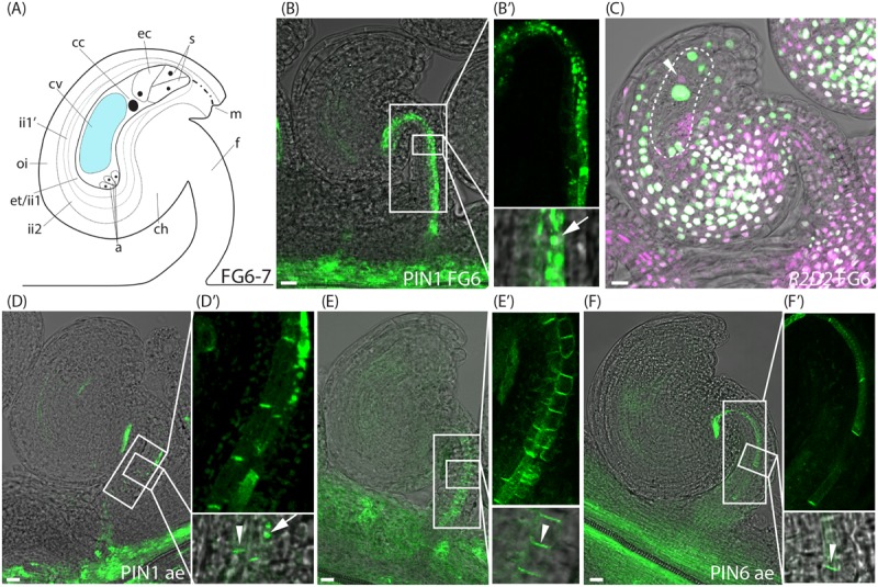
FIGURE 3.

Auxin sensing and transport in ovules at the pre-anthesis stage and after emasculation. (A) Schematic overview of an ovule at stage FG6-7. a, antipodal cells; cc, central cell; ch, chalazal domain; cv, central vacuole; ec, egg cell; et, endothelium; f, funiculus; ii, inner integuments; m, micropylar end; oi, outer integuments; s, synergid cells. (B,C) Merged confocal and transmitted light images of stage FG6 PIN1pro:PIN1-GFP (B,B′) and R2D2 (C) ovules just before anthesis. (B′) Shows higher magnifications of the boxed areas in (B), arrow indicates internal PIN1 localization. Dotted area in (C) indicates the female gametophyte, arrowhead in (C) indicates DII depletion in the egg cell. (D–F) Merged confocal and transmitted light images of PIN1pro:PIN1-GFP (D,D′), PIN3pro:PIN3-GFP (E,E′), PIN6pro:PIN6-GFP (F,F′) ovules after emasculation. (D′–F′) show higher magnifications of the boxed areas in (D–F), respectively. Arrowheads indicate basal PIN1, PIN3, and PIN6 localization, respectively, arrow indicates internal PIN1 localization. Bars = 10 μm. All images are representatives of at least 10 independent samples.
