FIGURE 4.
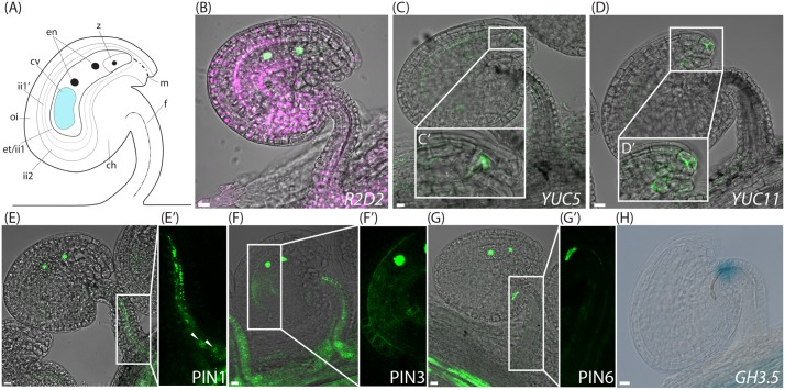
Auxin sensing, biosynthesis, transport and conjugation in ovules post-fertilization. (A) Schematic overview of an ovule after the first nuclear division of the central cell shortly after fertilization, ch, chalazal domain; cv, central vacuole; en, endosperm nuclei; et, endothelium; f, funiculus; ii, inner integuments; m, micropylar end; oi, outer integuments; z; zygote. (B,G) Merged confocal and transmitted light images of R2D2 activity (B), YUC5pro:3xGFP expression in the developing endosperm and in the micropylar end of the inner integument tip cells (C,C′), YUC11pro:3xGFP expression in the micropylar end of the inner integuments (D,D′), PIN1pro:PIN1-GFP expression in the funiculus (E,E′), PIN3pro:PIN3-GFP expression in the funiculus and in the inner integuments (F,F′), PIN6pro:PIN6-GFP expression (G,G′) in ovules at the two nucleate endosperm stage post-fertilization. (H) DIC image of a GUS stained GH3.5pro:GUS ovule at the two nucleate endosperm stage post-fertilization showing GH3.5 expression in the upper part of the funiculus. (C′–G′) Show higher magnifications of the boxed areas in (C–G), respectively. Arrowheads in (E′) indicate basal PIN1 localization. Bars = 10 μm. All images are representatives of at least 10 independent samples.

