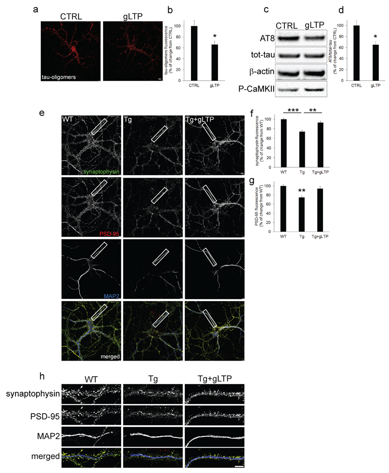
Figure 2. Synaptic activation reduced pathological Tau, and restored synaptophysin and PSD-95 to wild-type levels in PS19 cultured neurons.
a, b gLTP reduced levels of Tau oligomers (34±6%) compared to control treated (CTRL) Tg neurons, as quantified by confocal immunofluorescence (n=5; two-tailed unpaired t-test, *p<0.05; scale bar: 7.5μm). c, d Western blot analyses demonstrated a reduction (25±5%) of AT8 in gLTP compared to CTRL Tg neurons (n=5; two-tailed unpaired t-test, *p<0.05). e, h gLTP restored levels of synaptophysin (green) and PSD-95 (red) back to WT levels compared with CTRL Tg neurons, as quantified in f (n=5; one-way ANOVA test, p=0.0018; WT vs Tg ***p<0.001; Tg vs Tg+gLTP **p<0.01) and g (n=5; one-way ANOVA test, p=0.0068; WT vs Tg **p<0.01; Tg vs Tg+gLTP **p<0.01), respectively (scale bars: 7.5μm). “n” refers to a set of cultured neurons prepared from one mouse embryo. Three preparations of neurons were required and experiments were repeated accordingly.

