
Fig.6.
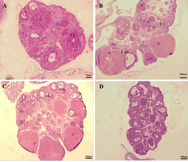
Representation of histological study on immature female rat ovaries. A. Ovaries from the control (magnification: ×10, scale bar=200 μm), B. Treated with 100 mg/kg, C. 200 mg/kg royal jelly (RJ), and D. 400 mg/kg of RJ. The figures show primary, secondary and graffian follicles and also observed the corpora lutea (magnification: ×4, scale bar=100μm). Primary follicles (F I), the secondary follicles (F II), the antral follicles (F III) and Corpora lutea (X).