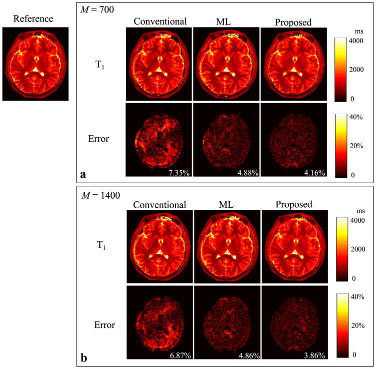
Fig. 3.
Reconstructed T1 maps for the slice 1 from the conventional MRF reconstruction, the ML reconstruction, and the proposed method (L = 8) with two acquisition lengths (i.e., M = 700 and 1400). a: Reconstructed T1 maps and corresponding error maps from the acquisition length M = 700. b: Reconstructed T1 maps and corresponding error maps from the acquisition length M = 1400. Note that the overall NRMSE is labeled at the lower right corner of each error map, and that the regions associated with the background, skull, scalp, and CSF are not included into the NRMSE calculation, and are set to be zero in each error map.

