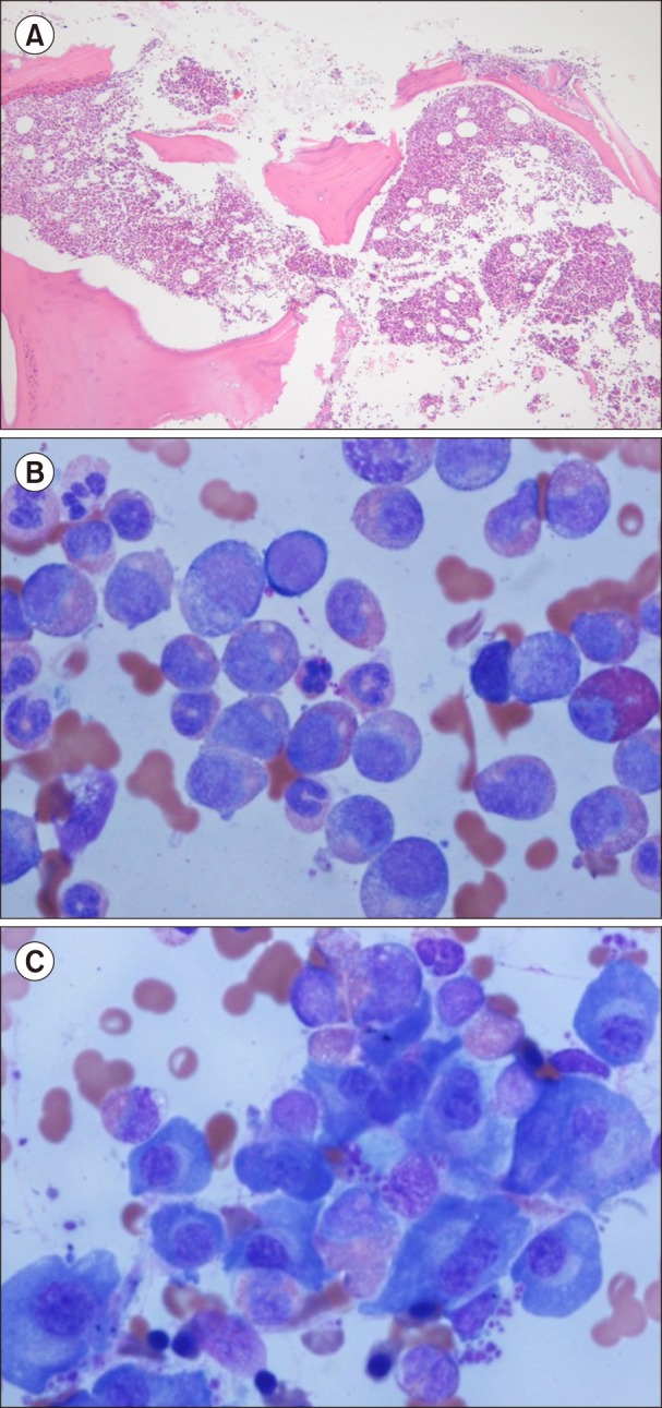
Fig. 2. Bone marrow biopsy and aspiration at diagnosis. (A) Bone marrow biopsy shows increased cellularity up to 90% (H&E, ×100). (B) Bone marrow aspiration shows myeloid hyperplasia with increased eosinophils (Wright-Giemsa staining, ×1,000). (C) Bone marrow aspiration shows increased mature forms of plasma cells (Wright-Giemsa staining, ×1,000).

