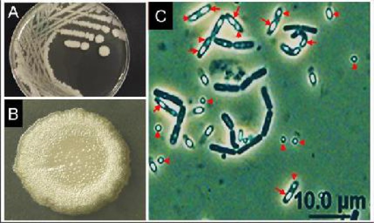
Fig. 2.
Photomicrographs showing morphology of the native Bt-55 isolate seeded in nutrient-supplemented agar media. A–B: colonies and a single magnified colony showing white, raised-centrally, nearly-circular, and glossy colony morphology with fine irregular margins similar to that of Bti-H14. (Photos A and B by AMA). C: Phase-contrast microscopy (×1000) illustrating the parasporal crystal (arrow heads) and bacterial spores (arrows) which appear brighter

