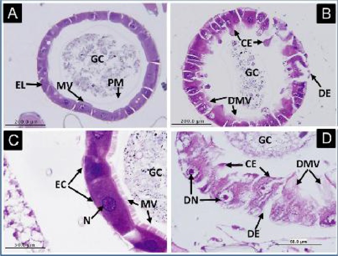
Fig. 3.
Histopathological impact of HS1 toxin-complexes on the midgut epithelia of Cx. pipiens larvae, 24h post-treatment. Figs. A and C represent cross sections in midguts of untreated larvae, showing normal gut epithelial layer (EL) with healthy, normal epithelial cells (EC), peritrophic membrane (PM), microvilli (MV), nuclei (N), and nutritional gut contents (GC) filling the gut lumen. Figs. B and D represent cross sections in midguts of treated larvae, showing affected gut epithelial layer, with cytoplasmic extensions (CE), degraded microvilli (DMV), degenerated epithelial cells (DE)

