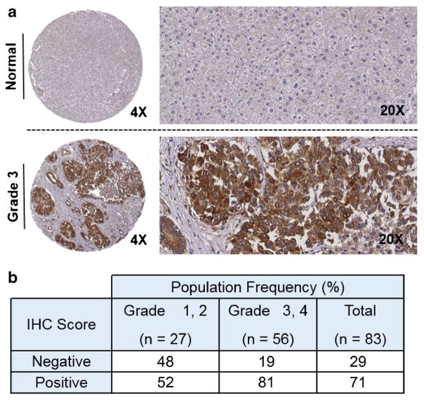
Fig. 3.
Tumor microarray analysis of SLC7A11 expression levels by immunohistochemistry. a Typical level of staining for non-tumor liver, showing minimal immunoreactivity. In contrast, grade 3 HCC exhibits robust staining, with SLC7A11 immunoreactivity confined to tumor cells. Transporter localization localizes to the membrane, as expected. Magnifications (×4 and ×20) shown. b Statistical analysis of 83 tissue specimens from the Vanderbilt HCC cohort (p = 0.006).

