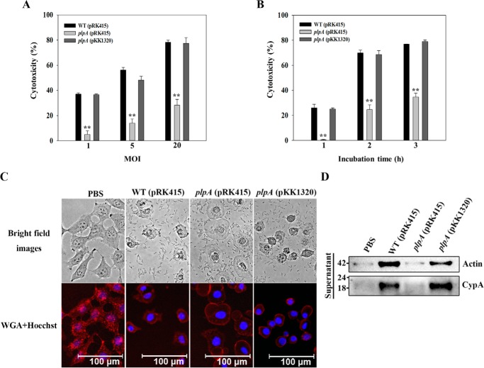
Figure 3.
VvPlpA is essential for necrotic cell death in tissue culture. A and B, INT-407 cells were infected with the V. vulnificus strains at various MOIs for 2 h (A) or at an MOI of 10 for various incubation times (B). The cytotoxicity was expressed using the total LDH activity of the cells completely lysed by 1% Triton X-100 as 100%. Error bars, S.D. Statistical significance was determined by Student's t test. **, p < 0.005 relative to groups infected with the wild type at each MOI or incubation time. C, INT-407 cells were infected with the V. vulnificus strains at an MOI of 10 for 1.5 h as indicated, stained using Texas Red®-X–conjugated WGA (for membrane, red) and Hoechst 33342 (for nucleus, blue), and then photographed using a fluorescence microscope. D, INT-407 cells were infected with the V. vulnificus strains at an MOI of 10 for 2 h, and actin and CypA released in the culture supernatants were analyzed by Western blot analysis. Molecular size markers (GenDEPOT, Barker, TX) are shown in kDa. WT (pRK415), wild type; plpA (pRK415), plpA mutant; plpA (pKK1320), complemented strain; PBS, control (uninfected).

