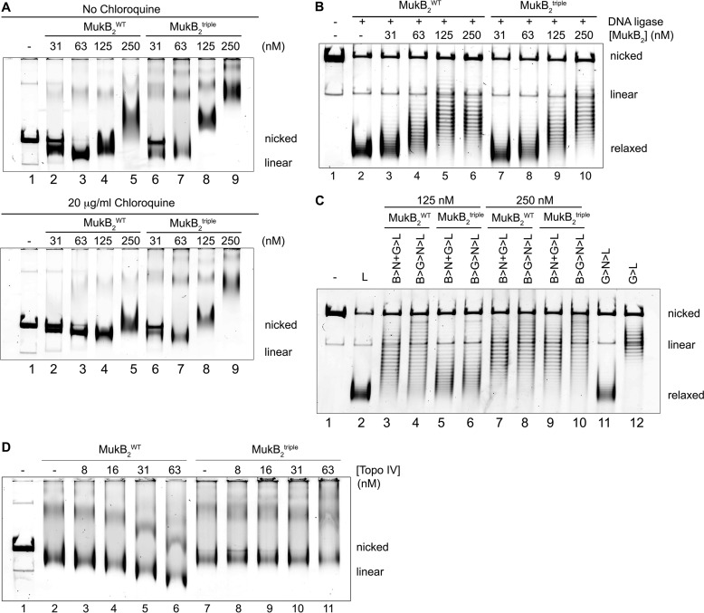
Figure 5.
MukB D697K/D745K/E753K variant condenses DNA but does not form maximally compacted DNA with Topo IV. A, DNA condensation by MukBtriple and wild-type proteins. The indicated concentrations of either MukBtriple or wild-type MukB were incubated with nicked DNA for 5 min at 37 °C, and the protein-DNA complexes formed were analyzed by gel electrophoresis either in the absence (upper panel) or presence (lower panel) of chloroquine as described in the legend to Fig. 1A. B, protection of negative supercoils by MukBtriple and wild-type proteins. The indicated concentrations of either MukBtriple or wild-type MukB were incubated with nicked DNA, E. coli DNA ligase, and NAD for 30 min at 37 °C. The samples were deproteinized, and the DNA was analyzed by electrophoresis through a 0.8% agarose gel in the presence of 10 μg/ml chloroquine in the gel and running buffer. C, loop stabilization by MukBtriple and wild-type proteins. MukB proteins were incubated with the nicked DNA substrate, 2 mm ATP, and 20 nm DNA gyrase for 30 min at 37 °C. Novobiocin was added to 10 μm, and the incubation was continued for 5 min. Bacteriophage T4 DNA ligase was then added, and the incubation was continued for 30 min. The products were deproteinized before analysis by electrophoresis through agarose gels containing 10 μg/ml chloroquine in both the gel and the running buffer. Relaxed, the nicked DNA sealed by DNA ligase to give a closed DNA ring that does not contain supercoils. The order of addition of components is outlined at the top of the gel. B, MukB; N, novobiocin; G, DNA gyrase; and L, DNA ligase. D, MukBtriple cannot form maximally condensed DNA with Topo IV. The indicated concentrations of Topo IV and either MukB or MukBtriple were incubated with the nicked DNA for 5 min at 37 °C, and the protein-DNA complexes formed were analyzed by gel electrophoresis as described in the legend to Fig. 1A.

