
Figure 11.
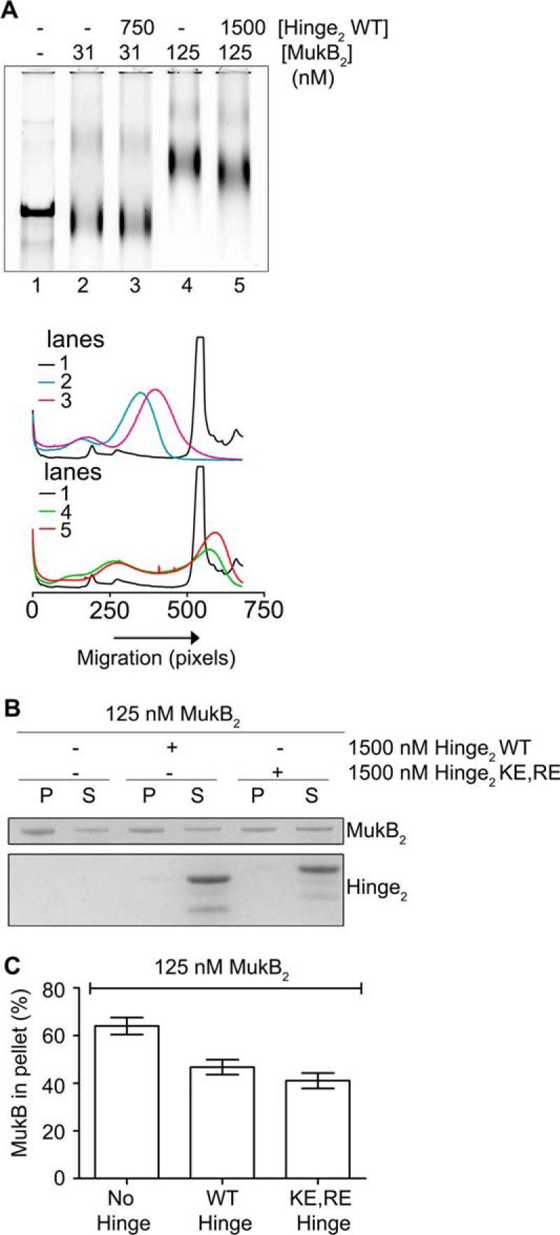
Hinge domain fragment destabilizes bound MukB. A, tailed, nicked DNA substrate is equivalent to the nicked DNA substrate. Top panel, MukB in the presence or absence of the wild-type hinge domain fragment was incubated with the biotinylated, tailed, nicked DNA substrate for 5 min at 37 °C and then analyzed by agarose gel electrophoresis. Bottom panel, densitometric tracings of the lanes of the gel shown in the top panel. B and C, hinge domain fragment destabilizes bound MukB. MukB was incubated on a rotator with tailed, nicked DNA substrate that had been bound to magnetic beads for 5 min at 37 °C. Either wild-type or KE,RE hinge domain fragment was then added, and the incubation continued for 30 min. The beads were then pulled down on a magnet, and the supernatant was removed. The protein present in the pellet (P) and supernatant (S) fractions were then assayed by SDS-PAGE. B, example of the SDS gel. C, distribution of MukB in the pellet and supernatant. The mean and standard deviation is shown for three independent experiments.