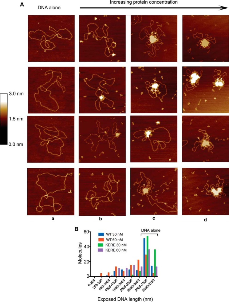
Figure 3.
Progressive DNA condensation of DNA by MukB as visualized by scanning force microscopy. A, protein-DNA complexes formed with the nicked DNA substrate either in the absence of MukB (column a) or in the presence of 15 (column b), 30 (column c), or 60 nm MukB (column d) were imaged in the SFM as described under “Experimental procedures.” Images shown are for wild-type MukB. Images for MukB KE,RE were identical in appearance. B, extent of exposed DNA in the MukB-DNA complexes, measured as described under “Experimental procedures,” is presented as a function of MukB concentration. WT, wild type; KERE, MukB K761E/R765E protein variant. A total of 100 molecules was measured from three independent experiments.

