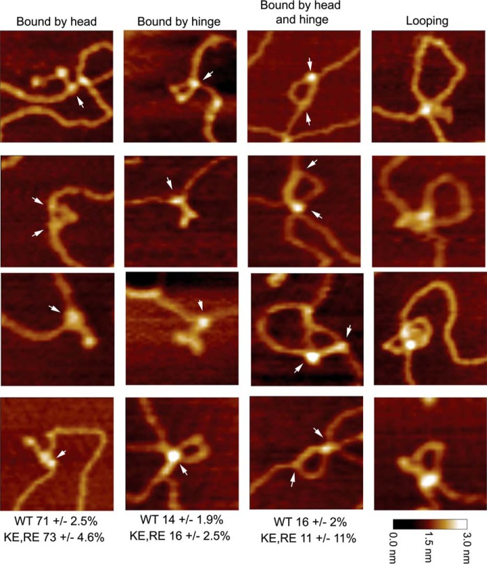
Figure 4.
Modes of MukB binding to DNA as visualized by scanning force microscopy. One hundred molecules were counted from three independent experiments where MukB at either 7.5 or 15 nm was bound to the nicked DNA substrate, and the modes of binding were characterized as either MukB bound to the DNA by a head domain, MukB bound to the DNA by the hinge domain, or MukB bound to the DNA by both the hinge and head domains. The fraction of molecules bound in each mode for the wild type and the MukB KE,RE variant is given below each column. Images where the DNA is looped with MukB bound at the apex are also shown. Images shown are for wild-type MukB. Images for MukB KE,RE were identical in appearance. Arrows indicate points of contact between MukB and the DNA.

