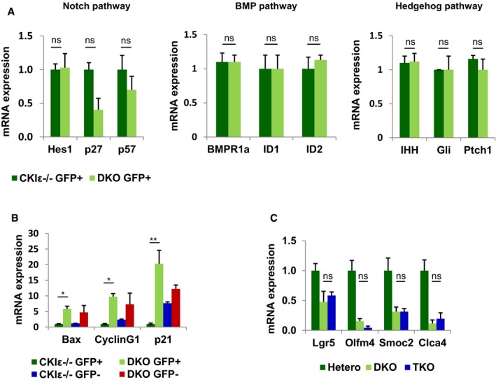
Figure EV4. Extinction of DKO ISCs is not mediated by Notch signaling downregulation or p53 activation.

- qRT–PCR analysis of Notch, BMP, and Hedgehog target genes in sorted GFP+ cells isolated from crypts of CKIε−/−‐VillinCreER‐Lgr5CreER (CKIε−/−) and DKO‐VillinCreER‐Lgr5CreER mice (DKO) 2.5 days after KO induction. Data represent mean of three independent sorting experiments, each with a pool of six mice; t‐test was performed, ns = non‐significant. Error bars indicate SEM.
- qRT–PCR analysis of p53 targets in sorted GFP+ and GFP− cells isolated from crypts of CKIε−/−‐VillinCreER‐Lgr5CreER (CKIε−/−) and DKO‐VillinCreER‐Lgr5CreER (DKO), 2.5 days after KO induction. Data represent mean of three independent sorting experiments, each with a pool of six mice; t‐test was performed, *P < 0.05 **P < 0.01. Error bars indicate SEM.
- qRT–PCR analysis of ISC markers in IECs isolated from CKIδ hetero (hetero; n = 3), DKO (n = 3) and TKO (n = 3) mice (mean ± SEM), 5 days after KO induction; t‐test was performed, ns = non‐significant.
