
Figure 2. In vivo interaction between release factors and UPF proteins.
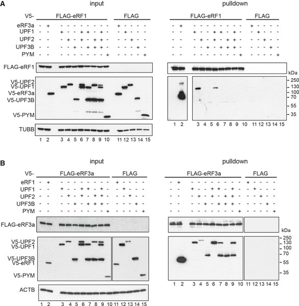
- Co‐immunoprecipitation from RNaseA‐treated lysates of HeLa cells transfected with FLAG‐eRF1 (lanes 1–10) or unfused FLAG (lanes 11–15) and V5‐eRF3a, V5‐UPF1, V5‐UPF2, V5‐UPF3B or V5‐PYM. Co‐precipitated proteins were detected using an anti‐V5 antibody. Lysate used for the immunoprecipitations was loaded in the input lanes (left). Re‐probing with anti‐TUBB antibody served as loading control.
- Co‐IP experiment as in (A) with FLAG‐eRF3a. Re‐probing with anti‐ACTB served as loading control.