
Figure 4. UPF3B can directly interact with UPF1.
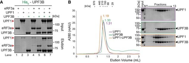
- In vitro pulldown of eRF3a, UPF1 or both with His‐UPF3B. Protein mixtures before loading onto the beads (input) or after elution (eluate) were separated by SDS–PAGE. Representative of four independent experiments.
- Left: SEC elution profile of UPF1 (purple), UPF3B (green) or both (orange). Right: SDS–PAGE analysis of eluate fractions. Representative of two independent experiments. Since the experiments described in Figs 3F and 4B were performed in parallel, the same UPF3B SEC elution profile (green) and the corresponding SDS–PAGE analysis served as control for both experiments. More UPF3B SEC experiments are depicted in Figs 3G, 6B and EV3E.
Source data are available online for this figure.