
Figure 6. UPF3B binds RNA and ribosomes.
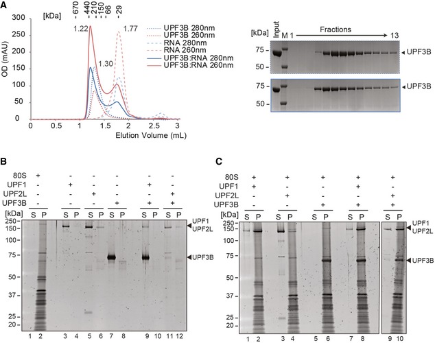
- Left panel: SEC elution profile of UPF3B (dotted lines), RNA (dashed lines) and the mix (solid lines). Optical density was recorded at 280 nm (blue) and at 260 nm (red). 40 μM of UPF3B or RNA oligonucleotide (24mer) or both was loaded onto a Superdex 200 column. The elution volumes are indicated next to the curves. Right panel: SDS–PAGE analysis of eluate fractions.
- Sucrose cushion co‐sedimentation analysis of either ribosomes (lanes 1, 2) or of UPF1, UPF2L or UPF3B or of combinations as indicated. After ultracentrifugation, the supernatant (S) and pellet (P) fractions were analysed by SDS–PAGE.
- Sucrose cushion co‐sedimentation analysis of UPF1, UPF2L or UPF3B as in (B) but in the presence of 80S ribosomes.