
Fig. 4.
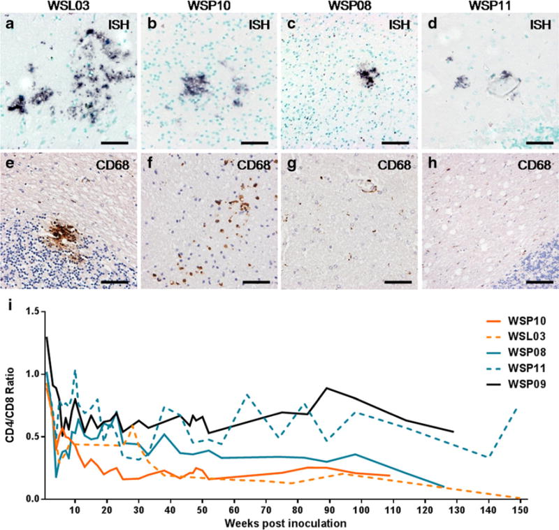
Variant neuropathological changes and CD4/CD8 ratio over time in CP macaques a–d. ISH for SIV RNA (BCIP/NBT; indigo) in brains of CP macaques. More SIV-expressing cells presented in the brain of WSL03 (a, temporal cortex) and WSP10 (b, occipital cortex) comparing to WSP08 (c, occipital cortex) and WSP11 (d, cerebellum). e–h. Distribution of CD68+ macrophages (DAB; brown) in brains of CP macaques with SIVmac239 infection. e. CD68+ macrophage accumulation was seen in the cerebellum of WSL03. f. CD68+ macrophages scattered in the brain of WSP10. g–h. IHC staining of CD68 was not significant in the brain of WSP08 (g) and WSP11 (h), while ISH for SIV-RNA was positive (c and d). i. CD4/CD8 ratio in 5 CP macaques with a neuropathological diagnosis. Scale bars: 50 μm