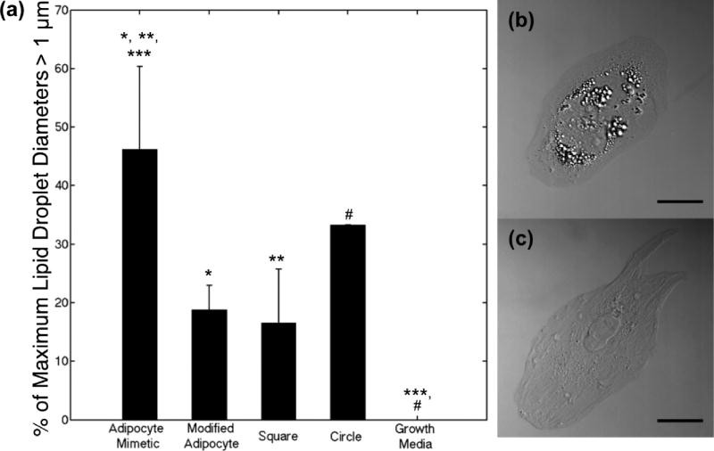
Figure 6.
Lipid droplet analysis of HMSCs following 7 days in culture. (a) Percentage of HMSCs displaying lipid droplets with maximum diameter greater than 1 µm on adipocyte mimetic (31 cells), modified adipocyte (33 cells), square (23 cells), and circle (33 cells) patterns, as well as cells cultured in growth media (no patterns, 32 cells). Results for at least 3 samples per pattern type are shown. Data are shown as mean ± standard deviation; significance was calculated using one-way ANOVA with a Tukey posthoc analysis (p < 0.05: adipocyte mimetic compared to modified adipocyte (*), square (**), and growth media (***); circle compared to growth media (#)). Representative DIC image of HMSC on an (b) adipocyte mimetic and a (c) modified adipocyte fibronectin pattern (scale bar = 25 µm).

