
Fig. 2. Disassembly of myofibrils in cultured single adult cardiomyocytes.
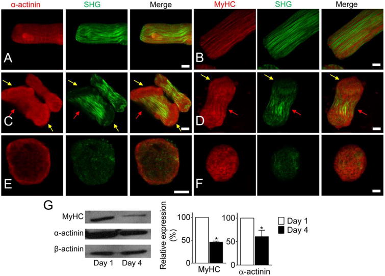
(A-B) LRS cardiomyocytes with clearly striated α-actinin, MyHC, and myofibril (SHG). (C-D) Myofibrils were disassembled from the cell ends in peanut-shape cells. α-actinin accumulated at the end of cells (yellow arrows), striated Z-discs disappeared, and myosin filaments broke up. But MyHC molecules were still detected at the cellular regions. The striated structure was still visible at the middle region (red arrows). (E-F) myofibrils had totally disassembled in the RS cardiomyocytes. α-actinin accumulated at the cell periphery and MyHC randomly aggregated in the cell. (G) Western blot and semi-quantitative data of MyHC and α-actinin in cardiomyocytes cultured Day 1 and Day 4, respectively. * P < 0.05, compared with Day 1. Scale bar: 10 μm.