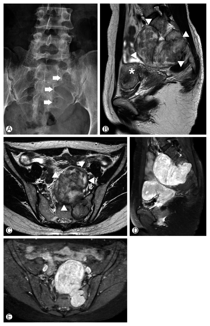
Fig. 1.
Preoperative radiographic images of a mass lesion located in the presacral space. (A) Anterior posterior plain radiograph of the sacrum demonstrating the widened neural foramen at the left S1–3 level (arrows). (B, C) Sagittal and axial T2-weighted magnetic resonance (MR) images showing a heterogeneous iso/high intensity mass (arrowheads) at the presacral area displacing the uterus (asterisk). (D, E) Sagittal and axial T1-weighted postcontrast MR images showing a well-enhanced mass compressing the pelvic organs.

