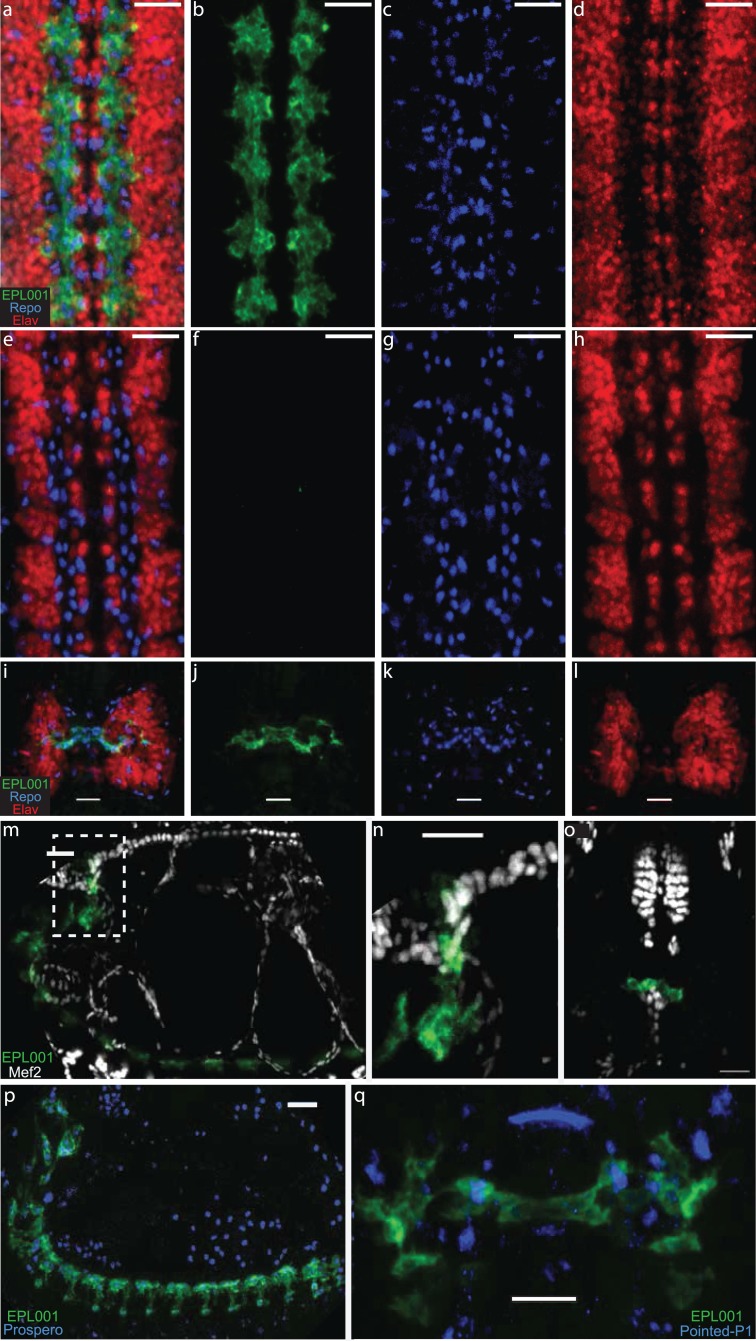
Figure 4. Fruit Fly IHC.
Goat anti-EPL001 antiserum G530 used throughout, at a dilution of 1/100, staining in green; Repo marker in blue and Elav marker in red. Scale bars 20 μm throughout. (A) Fruit fly embryo ventral nerve cord, viewed from ventral side, merged image of anti-EPL001 antibody, Repo and Elav. (B) Anti-EPL001 antibody channel only. (C) Repo channel only. (D) Elav channel only. (E) Preabsorption control with EPL001 peptide added, merged image (as A). (F) Anti-EPL001 antibody channel only. (G) Repo channel only. (H) Elav channel only. (I) Brain, viewed from dorsal side of embryo, merged image of three stains (as A). (J) Anti-EPL001 antibody channel only. (K) Repo channel only. (L) Elav channel only. (M) Lateral view of embryo, with anti-EPL001 antibody in green and Mef2, which stains heart and mesoderm, in white. (N) Zoom of inset dotted area of M. (O) Ventral view of embryo, staining as M, with heart seen abutting the anti-EPL001 antibody staining of brain and pharynx. (P) Lateral view of embryo, staining with anti-EPL001 antibody and Prospero, a lateral glia marker, in blue. (Q) Brain region, dorsal view, showing partial co-localisation of anti-EPL001 antibody with Pointed P1, a stomatogastric marker, in blue.

