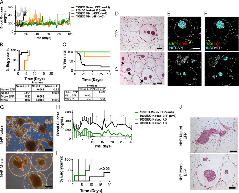
FIGURE 2.

Effects of transplantation site on the outcome of islet allografts encapsulated in optimized ALG microcapsules (Micro) without immunosuppression. The free-floating intraperitoneal (IP) site is compared with the confined and vascularized EFP site. A, Blood glucose of STZ-induced diabetic C57BL/6 mice transplanted with 750 IEQ naked in the EFP (black, n = 15) or IP (grey, n = 8) sites or 750 IEQ microencapsulated (Micro) in the EFP (green, n = 7) or IP (orange, n = 5) sites; all islets from fully MHC-mismatched BALB/c mice donors. B, Percentage of mice that reversed diabetes after transplantation. C, Percentage survival of allografts that reversed diabetes after transplantation. Tables below graphs indicate P values. D-F, Histological evaluation of EFP grafts and of capsules retrieved from the IP site by intraperitoneal lavage, fixed in formalin, embedded in paraffin, and thin sliced (5 μm). Shown are grafts that reversed diabetes and maintained euglycemia for more than 100 days. In H&E-stained sections (D) arrows point at areas of islet central necrosis. Scale bars, 100 μm. Confocal images: host vessels (CD31+, red), macrophages (MAC2+, green) and beta cells (INS+, cyan) are shown in panel (E); T cells (CD3+, red), B cells (B220+, green) and beta cells (INS+, cyan) are shown in panel (F). Nuclei are counterstained with 4′,6-diamidino-2-phenylindole (DAPI) (grey). Scale bar 150 μm; (G) Phase contrast images of baboon islets encapsulated in Micro capsules fabricated with optimized fabrication parameters (Table 1, bold) and loading density of 15 k IEQ/mL and compared to Naked islets. Scale bars, 200 μm; (H) Blood glucose of STZ-induced diabetic NOD-scid mice transplanted with 1000 IEQ naked (black, n = 5) or 750 IEQ microencapsulated (Micro, green, n = 4) islets in the EFP and compared to 1000 IEQ (light grey) and to 2000 IEQ (dark grey) naked islets transplanted in the kidney capsule (KD) controls; all islets from baboon nonhuman primate donors. I, Percentage of mice that reversed diabetes after transplantation of baboon islets. J, Histological evaluation of EFP grafts of naked versus Micro encapsulated in the EFP site analyzed 30 days after transplantation in diabetic NOD-scid mice. Scale bars 200 μm.
