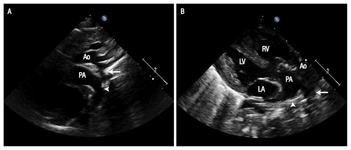
Figure 2.
Echocardiograms of a six-week-old female infant in the (A) suprasternal and (B) parasternal long axis views showing coarctation (arrow) and the continuation of the arterial duct (arrowhead) into the descending aorta. Note the anterior aorta arising from the right ventricle and the posterior pulmonary artery.
Ao = ascending aorta; PA = pulmonary artery; LV = left ventricle; RV = right ventricle; LA = left atrium.

