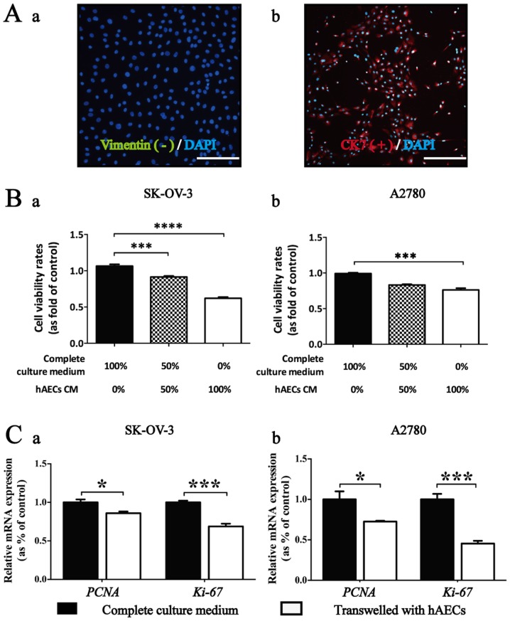
Figure 1.
hAECs inhibit proliferation of EOC cells in vitro in a paracrine manner. (A) hAECs was negative for mesenchymal marker vimentin (green) and positive for epithelial marker CK-7 (red) by using immunofluorescence. DAPI (blue) staining showed the nuclei (scale bar, 100 µm). (B) CCK-8 cell viability assay used to test the effects of hAEC-CM in different concentrations on the viability of EOC cells at 48 h (n=6; performed in triplicate). Ordinary ANOVA was used for statistical analysis. (C) Real-time PCR were used to test the effects of hAECs on the expression levels of PCNA and Ki-67 in EOC cells cultured in Transwell system for 48 h (n=3; performed in triplicate). Data are represented as means ± SEM. *p<0.05 and ***p<0.001.

