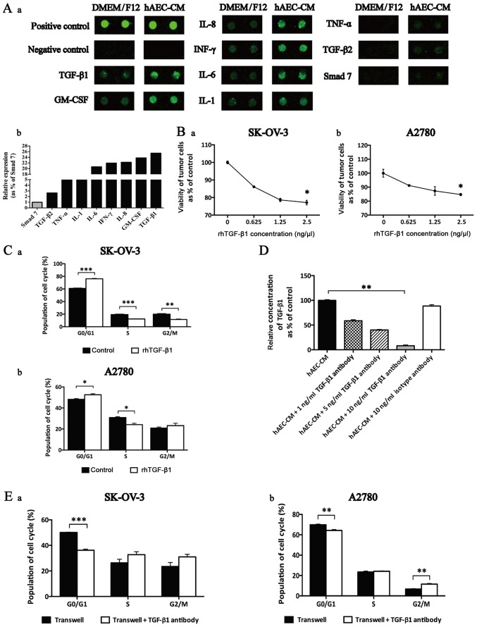
Figure 5.
TGF-β1 was enriched in hAEC-released cell cycle-regulatory cytokines and induced G0/G1 cell cycle arrest in EOC cells. (A) (a) Selective map of the human antibody array 1000. hAECs released multiple cell cycle-regulatory cytokines, including TGF-β1, GM-CSF, IL-8, IFN-γ, IL-6, IL-1, TNF-α, TGF-β2 and Smad 7. (b) The intensities of the signals were quantified by densitometry and the expression of Smad 7 was regarded as control. (B) CCK-8 cell viability assay used to test the effects of rhTGF-β1 in different concentrations on the viability of EOC cells at 48 h (n=6; performed in triplicate). Data were analyzed by Mann-Whitney U test. (C) After being treated with rhTGF-β1, cell cycle of EOC cells was analyzed by flow cytometry. (D) ELISA was used to test the efficiency of TGF-β1 antibody added to neutralize the function of hAEC-secreted TGF-β1. Data were analyzed by Kruskal-Wallis test. (E) After being treated with TGF-β1 antibody in the Transwell system, cell cycle of EOC cells was analyzed by flow cytometry. Data are presented as means ± SEM. *p<0.05, **p<0.01 and ***p<0.001.

