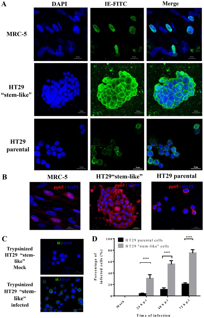
Figure 2.
The infection of HCMV laboratory strain AD169 in colorectal cancer-derived cells. (A) HCMV AD169 was used to infect 103 parental and stem-like HT29 and MRC-5 (control) cells at MOI of 5 on coverslips, followed by IE and (B) pp65 immunofluorescence. (C) IE-positive cells were observed after counter staining using a fluorescence microscope fitted with a camera. Five low magnification fields were counted for each condition. (D) The percentage of positive cells was calculated by dividing the number of IE-positive cells with the total number of nuclei, followed by multiplication with 100. ****p<0.0001 by two-way ANOVA and Tukey's multiple comparisons test.

