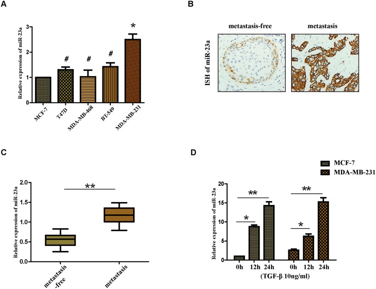
Figure 1. Expression of miR-23a in cell lines and human tissues and the treatment of TGF-β1 upregulates the expression of miR-23a in MCF-7 and MDA-MB-231 cell lines.
(A) Real-time PCR analysis of the expression of miR-23a in different breast cancer cell lines, normalized to GAPDH. (B) Analysis of miR-23a in situ hybridization(ISH) signal in breast cancer tissues. Representative images are shown (400 magnification). (C) Real-time PCR analysis of miR-23a expression in a group of 30 BC patients without lymph node metastasis and matched patients with lymph node metastasis. (D) Real-time PCR analysis of the expression of miR-23a in MCF-7 and MDA-MB-231 cells treated with 10ng/ml TGF-β1 for 12h or 24h. *P<0.05, **P<0.01.

