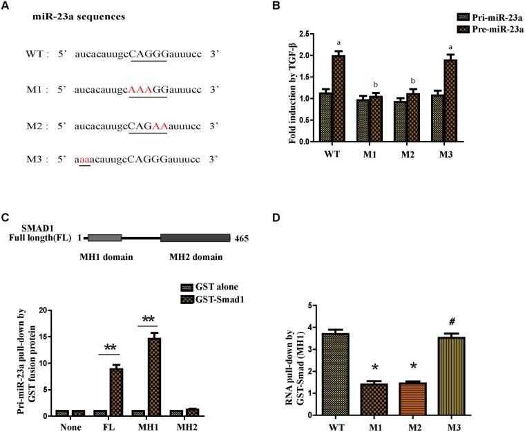
Figure 2. TGF-β1 regulates miR-23a post-transcriptionally and the R-SBE sequence is essential for the association of SMAD MH1 domain and miR-23a.
(A) Schematic diagram of pre-miR-23a wild-type and mutant sequences. Underlined characters indicate the R-SBE sequence found in pre-miR-23a and red highlighted characters indicate mutation introduced. (B) MCF-7 cells were transfected with human pri-miR-23a expression constructs, followed by treatment with or without 10ng/ml TGF-β1 for 2h and subjected to real-time PCR analysis using primers to detect exogenous pri-miR-23a or pre-miR-23a, normalized to GAPDH. Fold induction by the TGF-β1 relative to mock treated cells was displayed. Values labeled with different letters differed from one another (P<0.05). (C) The schematic diagram of domains of Smad1 protein (upper panel). In vitro transcribed wild type pri-miR-23a was mixed with indicated recombinant, sepharose bead-immobilized GST-fusion proteins. Associated RNA was eluted, and subjected to real-time PCR analysis to detect pri-miR-23a. The relative amount of pri-miR-23a pulled down with GST-Smad fusion proteins, normalized to the amount pulled down with GST alone is presented (lower panel). (D) In vitro transcribed pri-miR-23a constructs were mixed with recombinant GST-Smad1 (MH1) or GST alone and the relative amount of pri-miR-23a transcripts pulled down with GST-Smad1 (MH1) fusion protein was normalized to the amount pulled down with GST alone. *P<0.05, **P<0.01.

