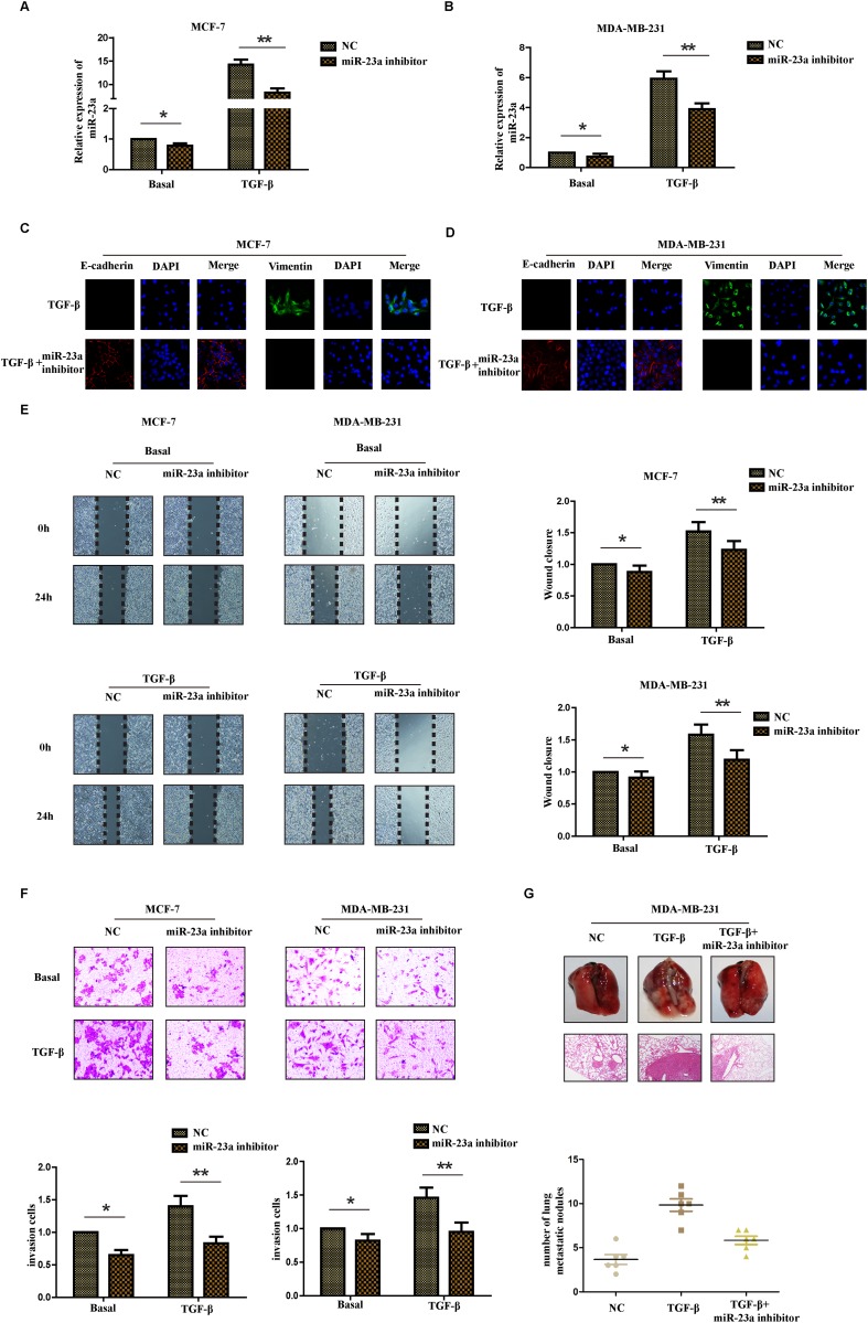
Figure 3. Inhibition of miR-23a suppresses the TGF-β1-induced EMT, the migration and invasion ability of breast cancer cells treated with TGF-β1.
Real-time PCR was used to confirm the transfection efficiency of miR-23a in MCF-7 (A) and MDA-MB-231 (B) cells treated with or without TGF-β1. (C and D) Representative IF images indicated that miR-23a had an effect on the expression of EMT genes in indicated cells treated with TGF-β1. (E) Representative micrograph images of wound healing assay of the indicated cells. Wound closures were photographed at 0h and 24h after wounding. (F) Invasion assays of the indicated cells transfected with NC or miR-23a inhibitor. (G) Tumor metastasis analysis. Representative images of lung tissues and micrographs of HE staining of metastatic tumor tissues (upper panel). The number of metastatic lung nodules in each group of nude mice (n= 6 per group, lower panel). *P<0.05, **P<0.01.

