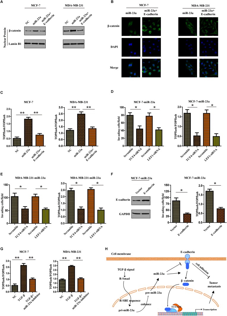
Figure 6. MiR-23a targets CDH1 to hyperactivate Wnt/β-catenin signaling and subsequently mediates the TGF-β1-induced EMT and tumor invasion in breast cancer.
(A) Nuclear fraction of indicated cells was analyzed by Western blot. Lamin B1 was used as a loading control. (B) Beta-catenin localization in indicated cells was detected by immunofluorescence staining. (C) Indicated cells were transfected with TOPflash and Renilla pRL-TK plasmids, and subjected to dual –luciferase assays 48h after transfection. Reporter activity was normalized to Renilla luciferase activity. (D and E) Transwell assays were used to detect the quantification of invading cells transfected with indicated siRNAs and luciferase-reported TCF/LEF transcriptional activity in indicated cells were examined. (F) Western blot analysis was used to confirm the transfection of constructs containing CDH1 ORF in miR-23a overexpressed MCF-7 cells (left panel). Quantification of invading cells transfected with indicated construct (middle panel) and luciferase-reported TCF/LEF transcriptional activity in indicated cells (right panel). (G) Luciferase-reported TCF/LEF transcriptional activity in NC, cells treated with TGF-β1 or miR-23a silenced-cells treated with TGF-β1. (H) Schematic representation of a model for the role of miR-23a in the TGF-β-induced tumor metastasis in breast cancer. *P<0.05, **P<0.01.

