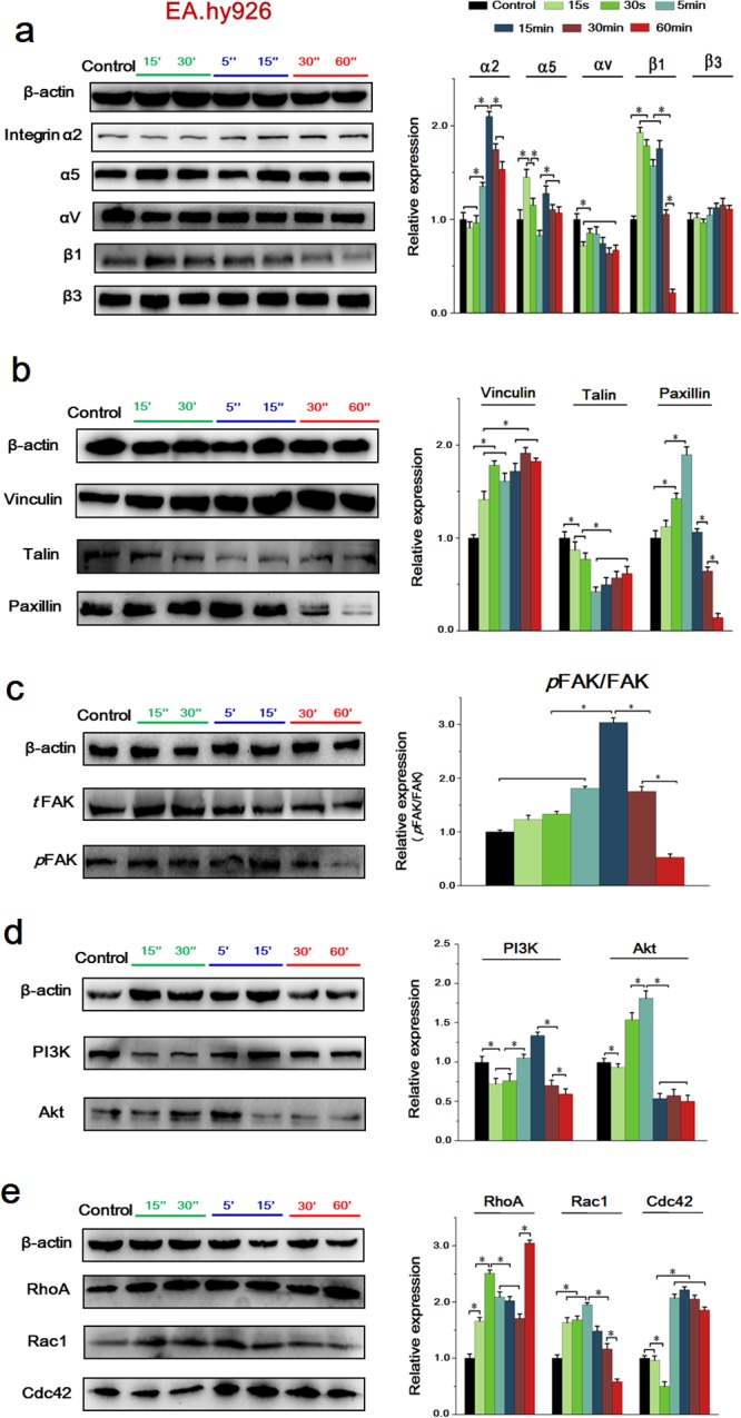
Figure 7. The expressions of key proteins and binding condition in Integrins-FAK-Rho GTPases signaling pathway of EA.hy926 cells.

(a) The expression of integrins, the expression levels were quantified by image analysis of the Western blot bands. The expression of β-actin in each group was used as intrinsic controls, and relative expressions of all of the proteins were calculated. (b) The expression of FAs, include Vinculin, Talin and Paxillin. The expression levels were quantified by image analysis of the Western blot bands. (c) The expression of total FAK and phosphorylated FAK, their expression levels were quantified based on image analysis of the Western blot bands. (d) The expression of PI3K and Akt, the expression levels were quantified based on image analysis of the Western blot bands. (e) The expression of Rho GTPases including RhoA, Rac1 and cdc42, as well as the expression levels were quantified by image analysis of the Western blot bands. Values represent the Mean±S.D from three independent experiments. *, P<0.05 denotes statistically significant difference compare with other groups.
